Sunday, December 31, 2023

New ultrathin materials can pull climate-warming CO2 from the air

Link to article

Click on images to enlarge.

The three most influential gases towards climate change are carbon dioxide (CO2), methane (CH4), nitrous oxide (N2O), and fluorinated gases. Of these, CO2 is present in the largest amount by far. Its contributions to the greenhouse effect from burning fossil fuels and other combustion processes were noted as far back as 1896 by physical chemist and Nobel Prize winner Svante Arrhenius. Methane and N2O have a greater potential to trap heat, but there is so little of them in the atmosphere that CO2's overall effect is stronger. Planting trees can help remove it because they use CO2 and sunlight to make their own food source via photosynthesis. Reducing the amount made from industry will also help, but regulations are not always easy to come by. What if there was a filter or type of "flypaper" to take CO2 out as fast as it is being made? There just might be.

Image from Pixabay

A special type of metallic material called a MAX phase was discovered naturally in the 1960s, but it took scientists until the 1990s to make them artificially. The name comes from how the three parts are labeled.

  • M = elements like the metals vanadium and titanium
  • A = elements like aluminum or silicon
  • X = carbon alone or carbon plus nitrogen

In the right combination, these form sheets that are one molecule thick for each of the three components. 

Image adapted from Science News Explores

The thinnest material in the world, however, is only one layer (molecule) thick. It is called graphene (from the word graphite because it is pure carbon). It was discovered in 1962 but ignored until 2004, when it was "rediscovered" and investigated. Graphene was found to be an important material for semiconductors, electronics, electric batteries, and composites. But because its chemistry was so simple, it had little other use.

Image of graphene sheet. Each dot represents a carbon atom (Wikipedia)

Because this single layer material has such different properties than the multi-layered MAX phases, scientists hoped to find something even more useful by removing the A element from a MAX phase in order to create a two-layered material they called a MXene (pronounced "max-een"). They did that with chemically etching away aluminum with hydrofluoric acid.

Diagram from Advanced Materials, 2011

Scientists like Michael Naguib at Tulane University have shown that MXenes can be made with many combinations of elements (about 50 now exist), and their properties can also be changed fairly easily. One chemical treatment gives a MXene the ability to soak up CO2. Raw MXenes are a black powder which can dissolve in hydrogen peroxide. That can be used as a paint or later dried in sheets. The amount of surface area from the powder is amazing. Just one gram (1/400 ounce) of MXene has the same surface area as a football field! That gives it a lot of filtering capacity in a small painted patch.

Microscopic photograph of the first MXene (Advanced Materials, 2011)

If MXene is mixed with cellulose (plant cell wall fiber) then an elastomer (rubberizing agent) and dried, it can be used in sheets to filter gas molecules, such as CO2 or methane from natural gas or chimney flue gases. Professor Per Persson, a materials scientist from the Swedish Linköping University, stated that such filters could be used on the top of industrial chimneys to prevent the exhaust from delivering CO2 into the atmosphere. 

Making a gas filtration MXene (figure adapted from Macromolecules, 2022)

One problem in the use of MXenes is the cost of the raw materials such as vanadium ($20/pound), titanium ($0.35/pound), and molybdenum ($5–$15/pound). Another is cleaning the CO2 off a MXene sheet after it has become saturated. Rinsing the sheets with hydrogen under high temperatures removes CO2, which can then be used as starting material for animal feed or methanol. The good news is that MXenes have many more uses:

  • Desalination (trapping energy of the sun to purify water by evaporation)
  • Waste water treatment (floating on water to evaporate it with 84% efficiency with sun energy)
  • Battery technology and energy storage (higher charging rates than lithium-ion batteries; charging batteries faster)
  • Nanogenerators (harvesting frictional energy from moving muscles to convert it into electric power)
  • Conductive coatings (retaining conductivity even under heavy stretching and bending)
  • Sensors and chemical noses (detecting ammonia and acetone, which are indicators of ulcers and diabetes, with lower traces than sensors now used in medical diagnostics)


Friday, December 29, 2023

A vibrating pill could help treat obesity, pig study finds

Link to article

Click on images to enlarge.

They say that weight loss is a matter of taking in fewer calories and burning as many as possible. A sort of supply and demand issue. But is the demand in one's head or stomach, and can that be controlled? Some diets purport stopping eating a meal when one feels 80% full, to allow for the body to catch up to itself and register a real feeling of fullness. But, many people can't control themselves, and so they continue to eat. It can be called making a pig out of oneself, but pigs might prove to be the solution to weight loss after all! Read on.

Image from Pixabay

Why do we feel full when we eat? Surrounding our stomach is a network of nerves called IGLEs (intraganglionic laminar endings). These are connected to nerve clusters called ganglia which run through two layers of smooth muscle outside of the stomach lining. IGLEs detect mechanical pressure from the stomach and send signals to the muscles to loosen or tighten, in order to allow food in or to squeeze it through to the small intestine. When they detect enough pressure, it suggests to the brain that the stomach is filled with food, and it then tells the body to stop.
IGLE nerve in a cross-section of stomach (adapted from Frontiers in Physiology)

If the goal is to force a person to eat less, then giving them a sensation of a full stomach is needed. Drugs are costly and potentially dangerous. Putting in a balloon was done as early as 1979. In 2017, researchers reviewed studies on the effects of inserting a balloon then filling it with saline (salt water) solution directly into the stomach. Lean people have a stomach volume of 1,000 milliliters (4 cups), and obese people's stomach has a volume of 1,920 milliliters (8 cups). So, various makers of stomach balloons allow for them to be inflated to 200, 400, 600, or 800 mL to take up stomach volume. Most balloons are inserted and filled under anesthesia, which adds another layer of complication and cost.
Stomach balloon insertion and inflation (Mayo Clinic)

The 2017 review found mixed results. Whether patients had diet treatment, and whether the balloon was a certain size both affected the results in a 3-month study.

  • balloon only (300 mL): 3.2 kg lost + 4.1 kg
  • balloon plus diet: 5.1 kg lost + 4.7 kg
  • diet only: 6.9 kg lost + 5.9 kg
  • no treatment: 0.6 kg lost + 2.5 kg

Researchers at Harvard University wondered whether stimulating the muscles around the stomach lining could be done and what effect that would have. Electrical stimulation was probably not a feasible technique. As far back as 1966, it was known that vibrations like those used in physical therapy massage, when applied to the body, can cause muscles to contract. So, Professor Shriya Srinivasan and a team of scientists at Harvard University developed a small device called a Vibrating Ingestible BioElectronic Stimulator (VIBES) pill which patients could swallow before eating. As the pill rested against the inside of the stomach, its vibrations might cause the brain to think stomach muscles were reacting to a volume of food in the stomach. The VIBES pill is 31 mm (1.2 inches) long and 9.8 mm (0.38 inches) wide.

VIBES pill inside the stomach of a pig (Science Advances 2023)

It has a motor and battery. The pill is coated with gelatin that holds down a pin; when stomach acid dissolves the gelatin, the pin moves to the on position to start the vibration. The vibrating waves trick the IGLEs nerves to send a false signal of an expanding stomach to the vagus nerve and then to the medulla in the brain. When the brain has the illusion that the stomach is full, it then reduces the amount of a "hunger hormone" sent to the body. 

Faking fullness with the VIBES pill (Science Advances, 2023)

Srinivasan's team confirmed what the VIBES pill did as follows. First, they inflated the pig's stomach with an endoscope to 30%, 60%, and 90% of its maximal volume. When they held those volumes for 3 minutes, they recorded electrical impulses from the vagus nerve. They compared those data with what vibrations at 60 Hz, 80 Hz, and 100 Hz did to the nerve. The results were virtually identical.

Top: results showing vagus nerve signal with endoscopic inflation of stomach
Bottom: results showing vagus nerve signal with vibration (from Science Advances, 2023)

Srinivasan and coworkers tested the pill on a dozen Yorkshire pigs 4-6 months old. After two weeks, and 108 meals, the experimental pigs ate less than those given a placebo. Pigs with VIBES pills ate 58.1% of food provided, while the control pigs ate 78.4% of food given to them, a significant difference.

Three pigs were tested further. They had the VIBES treatment, then none, then back on VIBES. Researchers measured the percentage of the meal that they ate during those times. How much they ate returned to normal right away without the pill (left graph, white area), which suggested eating behavior was dependent on the pill's vibrations. They also measured how much weight four pigs gained with and without the VIBES pill. The graph on the right shows less weight gained with VIBES treatment, matching the smaller amount of food eaten.

The concept of the VIBES pill is that people will take it before every meal, as the pigs did, and it will eventually find its way out of the body in the feces. No damage was seen to the stomach lining after Srinivasan's experiments were over, even though the pill is right at the maximum size allowed for humans to swallow it. The data from Harvard University will be used to set up human clinical trials. Pigs were chosen for this experiment because their stomach is similar to a human's, not just because of the stereotype of obese people eating like pigs.

Wednesday, December 27, 2023

Tiny living robots made from human cells surprise scientists

Link to article

Click on images to enlarge.

In 1920, Czech playwright Karel Čapek wrote a science-fiction play called "R.U.R.", which stood for Rossum's Universal Robots, and which was set in the year 2000. This was the first time the word robot had been used. These robots were more like androids, purely organic beings, instead of metallic walking computers. However, they looked like the tin man from the Wizard of Oz. Engineers have since been working on creating microscopic robotic machines to work at the cellular level. But in recent years, scientists have also gone back to the RUR days of organic robots. They are microscopic and composed of living cells. How does that work?

Robots in the play "R.U.R." (Wikipedia)

The benefit of such devices, if they were to exist today, is thought to be in drug delivery, surgery, diagnosis, monitoring of diseases like diabetes, and even cleaning the oceans. In 1959, Nobel Prize winner Richard Feynman even conceived of using tiny machines to more efficiently create new chemicals by directly manipulating atoms. 

What if it microscopic robots didn't require mechanical parts and were disposable? In 2020, teams at the  University of Vermont Advanced Computing Core and the Center for Regenerative and Developmental Biology at Tufts University developed organic microscopic robots. The Vermont group created computer models of potential cell clusters of skin and heart cells from the African frog Xenopus laevis. Skin cell properties were used to help build proposed structures in many shapes because skin cells are made to do that. Heart muscle cells flex, and their properties were added to the "evolutionary algorithm" in the software to determine various functions like movement. The idea was to see which shapes and configurations of these cells would theoretically work together in clusters to create functional organic robots.

100 computer models for functional cell clusters
(adapted from Proceedings of the National Academy of Sciences)
(red=heart cells, blue=skin cells)

After many computer trials, the most successful models were handed off to Tufts U. Scientists there then peeled apart cells from Xenopus embryos and put them in groups. The cells clumped together to form a cluster of many cells. Then, with tiny tweezers, the researchers poked and prodded the clusters into the shapes of the computer models. Each one took several hours to build, but eventually these became the living, moving "xenobots", named after the frog source. 

Computer model and colorized xenobot cell cluster (University of Vermont)

Just under a millimeter (0.04 inches) wide, they are about 3 times the size of a period in Times New Roman 12-point font. Tufts U researchers observed them move about in Petri dishes for days or weeks until their internal energy supplies ran out. Movement is caused by hair-like projections called cilia, which move like paddles. Some xenobots had holes in them designed to carry material, but others without holes spontaneously pushed pellets of material together as they moved.

Xenobot spinning, moving, joining other xenobots in a Petri dish (YouTube)

When xenobots were damaged even to the point of being cut nearly in half, they repaired themselves.

Clip from YouTube

But even more amazing is that these clumps of cells were able to reproduce. Instead of just splitting each cell in the cluster, they worked together to gather other cells in the area. Those new clusters became new xenobots that do the same at the "parents". See what the designer has to say:

From YouTube shorts

More recently, in November 2023,  the Tufts U researchers have created "anthrobots", which are cell cluster robots made from human cells, specifically adult lung cells. Instead of 3-4 days, these took 7 days before they showed movement. Also,  from 2,281 of these, about half consistently don't move at all even after 3 weeks, even though they have cilia. The thing is, since they were from lung tissue, the cilia formed on the inside of the clump like it was the lining of the trachea. By adjusting the chemical composition of the medium they lived on in the Petri dish, the researchers changed that to the outside.

Cilia shown in yellow (from Advanced Science)

Anthrobots tend to have 4 types of motion: circular, linear, curvilinear, and eclectic. Examples are shown below from the researchers paper.

Anthrobots come in 8 unique cluster shapes, and each is correlated with a certain amount of cilia. What's more, they follow a scratched line in the Petri dish, which might be useful in later medical research if these are used to follow tissue cuts for repair to take place. See below microscopic tracking of the scratch outlined in yellow and the anthrobot path in pink.

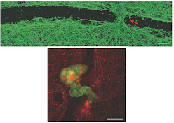
Taken to the next level, anthrobots have also been shown to fix damaged nerve tissue by forming "superbot" bridges. In the photos below, the top one shows a bridge formed in a cut. The bottom picture simply shows the superbot in red and nerve tissue in green.

Considering how important it is to repair nerve tissue, which normally is next to impossible, the usefulness of these organic robots is pretty clear.

Here's a nice 5-minute video that summarizes this blog article.


Sunday, December 24, 2023

Plants can call for help

Link to article

Plants are immobile; they can't run away from danger. But they have evolved various ways to protect themselves, for example, with sharp thorns or thick tree bark. Some plants even produce chemicals that are foul-tasting to invaders, which makes them less tasty and more likely to be avoided. Some make toxic substances, and others make chemicals that slow the growth rate of plant eaters. But some have found a way to call for help against predators. How do they do that?

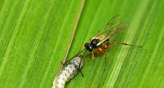
Wasp about to lay eggs inside a caterpillar attacking a plant (Science News Explores)

In the 1980s, University of Amsterdam scientists Marcel Dicke and Maurice Sabelis studied lima bean plants and their enemy, spider mites. When spider mites Tetranychus urticae attacked the plants, the damaged leaves in the top of the plant released 4 chemicals that are different from the ones released when leaves are mechanically damaged by other means. These four were specific "infochemicals" which female predatory mites (P-mites) use to locate and eat the spider mite and its eggs. The infochemicals were specific to attract the P-mites!

Orange predatory mite eating green spider mite (Univ. of CA Integrated Pest Management Program)

Swiss entomologist Thomas Degen in 2012 inoculated corn plant tops with an armyworm caterpillars using a spring-loaded "bazooka" and waited for them to feast on the corn. After that, he measured two things. Growing 6 types of corn in a lab, he placed a plastic bag over the top of the corn where the worms were eating, and using a pair of pumps, Degen could suck out air containing volatile chemicals (volatile means they evaporate easily into the surrounding air) that the plants made and replace it with fresh air. 

Collecting volatile chemicals from plants (Science News Explores)

Then, he analyzed the volatiles and found 36 chemicals were released. Six corn varieties each made different amounts, with the largest amount 15 times more concentrated than the lowest corn variety. And infested plants made more chemicals than controls with no infestations. He also noticed that the size of the worm larvae were smaller when the chemical concentration was higher. 

 
Bazooka inoculator design (Wiseman et al., 1980)
 
Inoculating plants with larvae using a "bazooka" (YouTube)

Degen noticed that in his early experiments in a lab (not the field), his infested plants made 9 times more chemicals than the non-infested controls in the lab. In the field, however, they made only 2.5 times more. One difference was that his lab plants were much younger (9-12 days vs 33-49 days). This got him and others to thinking why the younger plants would do that.

More recently, Lei Wang at the University of Bern in Switzerland has also studied corn to see what chemicals are released, and he saw a similar thing. Older plants don't make as much anti-predator warning chemicals. 

Chemicals from the left plant under attack trigger warning to right plant (modified from Science News Explores)

Nobody knows why young plant leaves do this better than old ones. They might be predisposed to doing it because the plant is still growing instead of at rest, and it may be more vulnerable then. New leaves are on the top of the plants, so the mechanism for chemical release might have evolved to take advantage of wind there. It's a pretty sensitive issue for growing plants, too. Ian Baldwin of the Max Planck Institute for Chemical Ecology in Germany showed as far back as 1983 that even if 7% of leaf damage was sustained by one plant, that was enough to send out a chemical signal in 1.5-2 days to warn neighboring plants. 

Middle plant signaling chemically to its neighbors that it is under attack. 
Note the spots on its leaves indicating insect larvae eating them. (From npr.org)

What is known is that some plants recognize chemicals in the saliva of an attacking insect larva, and that triggers the defensive release of the volatile plant chemicals. This is not limited to one species protecting its neighbors of the same species. Richard Karban (University of California - Davis) showed that damaging sagebrush can cause nearby tobacco plants to produce defensive chemicals. He toned down the name "plant communication" from one to another to a more casual term of "plant eavesdropping" to describe what may actually be happening. This might be because the signaling operates only within 50-100 cm (20-40 inches) from the plant. 

One benefit from these studies was expressed by Wang. Perhaps science could develop crops that are better at doing this and "smarter" to resist predators. 


For an historical perspective on learning how plants defend themselves, check out this link.

Monday, November 27, 2023

Charles Richard Drew: “Father of the Blood Bank”

Link to article

(Click images to enlarge.)

People don't often give a second thought to the process of donating blood or receiving it when needed. It's just always there in supply for routine and life-saving operations. But the history of blood transfusions and the nature of blood is a long one. And only 80 years ago did Dr. Charles Drew come up with a critical step in the storage of blood which has saved many lives.

From Wikipedia

William Harvey, an English doctor, discovered the circulatory system and published on it in 1628. Back then, the color of blood had been thought to indicate whether it was providing mere nutrients (purple blood) or some unique "vital factor", a life-giving principle from the lungs (red blood). Thirty years later, the Dutch scientist Jan Swammerdam looked at blood under a microscope and observed red blood cells for the first time. Nobody saw anything else until 1843, when Gabriel Andral, a French professor of medicine, and William Addison, an English doctor, separately noticed white blood cells. A year earlier, French physician Alfred Donné discovered platelets in blood, tinier than either red or white blood cells. These three made up the three solid components of blood as we know it today. The liquid part that carries these cells is called plasma.

Blood components after settling (Red Cross)

Charles Richard Drew was a Black American born on June 3, 1904 in Washington, D.C. It should be noted that he grew up in an interracial neighborhood in a middle-class family. At Dunbar High School, known as one of the best college prep schools in the country for blacks or whites, Drew was active in sports and earned letters in four of them. He joined the High School Cadet Corps there and became a captain. His goal at the time was not medicine but electrical engineering.

Drew's high school picture and description (Wikipedia)

He became interested in medicine at Amherst University, though, and graduated there in 1926. Drew decided to go teach biology and chemistry and be the football coach at Morgan University in Baltimore, Maryland for two years. That supplied him with enough money to attend medical school. For various reasons, he went to McGill University in Montreal instead of American schools. In 1933, he graduated second in his class while still playing sports there. As a resident then intern in 1933-1935 at Montréal General Hospital, Drew worked with bacteriology professor John Beattie, who studied exploring ways to treat shock with blood transfusions and other fluid replacement. 

Because places like the Mayo Clinic were not widely accepting Black physicians, he chose to work at Howard University College of Medicine (an all-Black institution) in Washington, DC in 1935. He was initially a pathology instructor, and then became a surgical instructor, and finally the chief surgical resident at its affiliated Freedmen's Hospital. The hospital was founded in 1862 and was known to be the first hospital to provide medical treatment to former slaves.

Drew teaching at Freedmen's Hospital (American Chemical Society)

From there, he gained surgical experience and moved to New York's Presbyterian Hospital, while simultaneously attending Columbia University beginning in 1938 for a doctorate in medical science. He was the first African American to earn such a degree there. At the hospital, he continued his interest in shock and transfusions. 

A short history is needed on transfusions in order to understand Drew's research and his later work.

  • 1665 The first recorded successful blood transfusion (England). Dogs to dogs.
  • 1667 More work on sheep, cows, dogs, horses, and goats. First transfusions from sheep to humans by Jean-Baptiste Denys (Denis). Because the lamb was a symbol of Jesus Christ and therefore would contain the purest blood, this was the reason it was chosen for humans. Initially a success, the patient then died, and a court case cleared Denys, but French and English governments put severe restrictions on transfusions after that for 150 years.
  • 1818 First successful transfusion of human blood (350 cc) by James Blundell to a patient.
  • 1873-1884 Americans attempt transfusing milk from cows, goats, and humans. Then they tried salt water (saline).
  • 1900-1905 Stitching together blood vessels for direct transfusions was perfected.

  • 1901 Austrian Karl Landsteiner discovers the first three human blood groups (A, B, O). Six years later, Ludvig Hektoen suggests that cross-matching blood between donors and patients would be safer, and Reuben Ottenberg and Albert Epstein perform the first blood transfusion using blood typing and cross-matching. Most physicians still thought that typing was not needed.
  • 1914 First indirect transfer of blood in a transfusion by Albert Hustin. Before this, patients and donors were connected directly. Indirect transfer means collecting the blood first into a container, then giving it to the patient.
Direct (left) and indirect (right) transfusion methods (indirect keeps blood warm in water bath)
  • 1915, 1916 Richard Weil and later Francis Rous and J.R.Turner add citrate or citrate-glucose to permits storage of blood for several days after collection.
  • 1932 First blood bank established (Leningrad) and in 1937 (Chicago)
  • 1936 John Elliot designed the first vacuum bottle for collecting blood
Elliot's vacuum bottle design (Transfusion, 2000)
  • 1939-1940 The Rh blood factor is discovered (you are either positive or negative).
  • 1940 Edwin Cohn developed a procedure to separate albumin, gamma globulin, and fibrinogen components from plasma.

Back to Charles Drew, with his newly minted doctoral degree awarded in 1940: "Banked Blood: A Study on Blood Preservation". What was it all about? Clearly, he had a lot of medical discoveries in recent years to fall back on.

Drew's dissertation mentions research from others in the 1920s suggesting how long blood could be kept under certain conditions that stabilized it (or "conserved" it). Those terms mean simply that care must be taken to ensure that during storage the red and white blood cells don't degenerate and that the blood doesn't clot. To quote from his dissertation:

p. 88 of Drew's dissertation

He looked into research on changes in blood's biochemistry and cell shape as well as immune properties, and he further spent 100 pages describing studies on preservation of blood, whether from placentas, cadavers, the aged, or freshly dead people. The blood chemistry changes during storage were explained in terms of illnesses that might benefit from transfusions. He even described the shape of containers and how they benefitted lengthy storage:

p. 198 of Drew's dissertation

He noted that the condition of blood was good up to 15 days at 3-5ºC (37-41ºF), compared to only 3 days at body temperature. Drew added that not only should blood be stored at that temperature, but that it should be chilled immediately after it was donated and kept "free from  mechanical shaking or movement" to reduce the risk of cellular damage. Ultimately, he determined that blood kept for 10 days under the physical and chemical conditions he outlined was as healthy as freshly drawn blood.

The final chapter in his dissertation described his proposal to establish a blood bank at his hospital. Survey results from four other hospitals in the U.S. (including Chicago) were used to incorporate valuable data in the creation of his own blood bank facility. His proposal was accepted. The result was a set of policies on administration, staff, blood drawing conditions of the hospital and patient, and sanitary conditions, and by the time he finished his dissertation, he had collected statistics (including health at blood drawing time and afterward) on the first 400 donors from August 9, 1939 to February 23, 1940!

Charles Drew, early 1940s

As mentioned already, blood is composed of liquid (plasma) with vital protein chemicals as well as the solid components of red and white cells and platelets. If they are together, it is called whole blood. Plasma was deemed more valuable than whole blood during wartime in many instances. Its loss from the body lowered blood pressure, and it was more important to restore that for the patient's stability than to provide red blood cells to boost oxygen-carrying capacity. Frozen plasma took 20 minutes to thaw, though, and it required special storage units. Freeze-dried plasma was more convenient in terms of storage and simply had to be reconstituted with sterile water before use.

British surgeons inspecting wartime blood, 1941 (American Chemical Society)

America was approached by Britain in World War II to help provide blood plasma for the expected casualties. In June 1940, Dr. Charles Drew was called upon to head up an effort to provide liquid plasma for British soldiers in the "Blood for Britain" campaign. With his guidance, the program scaled up his own hospital system to collect, process, and store plasma at nine hospitals. The first shipment went out in August. By the end in January 1941, Blood for Britain had collected 14,556 blood donations, and shipped over 5,000 liters of plasma with help from the American Red Cross. Drew was now considered the expert on blood donations and processing.

From February to April 1941, Drew worked again with the Red Cross to initiate a dried plasma program this time, which later became the National Blood Donor Service. 

Freeze-dried plasma (British Medical Journal, 1940)

It was Drew who introduced mobile collection units (later called "bloodmobiles.")

Drew's first bloodmobile, Feb. 1941 (American Chemical Society)

Despite his fame and success in assembling medical teams, the National Blood Donor Service became a sore point for Charles Drew. At the insistence of the Armed Forces, Blacks were not allowed to donate blood, and in retaliation, black news agents and the National Association for the Advancement of Colored People (NAACP) voiced their protests. A year later in 1942, the Red Cross changed its policy but insisted on separating blood from white populations. There was nothing scientific to suggest that Black blood was inferior or dangerous, but nothing could be done. A few months later, however, he resigned Drew himself, a light-skinned Black, was outraged and even mentioned this in 1944 in his acceptance speech for the Spingarn Medal, an honor from the NAACP for outstanding achievement:

"It is fundamentally wrong for any great nation to willfully discriminate against such a large group of its people. . . . One can say quite truthfully that on the battlefields nobody is very interested in where the plasma comes from when they are hurt. . . . It is unfortunate that such a worthwhile and scientific bit of work should have been hampered by such stupidity."

Charles Drew maintained a busy life thereafter. For example, he continued to serve as the chief surgeon at Freedmen's Hospital and as a professor at Howard University. He became the first African American examiner for the American Board of Surgery. He participated in Office of Civilian Defense (OCD) drills, created in 1941 by President Roosevelt. 

Practicing civilian defense in mock medical emergency for OCD, ~1942 (National Archives)

Drew dedicated himself to training and mentoring medical students and surgical residents, and raising standards in Black medical education. He was also an advocate against excluding Black physicians from local medical societies, medical specialty organizations, and the American Medical Association. Virginia State University and Amherst University both awarded him with honorary doctor of science degrees in 1945 and 1947. 

Tragically, at only 45 he died due to complication following a car accident on April 1, 1950 on his way to a medical conference in Alabama. An episode of the TV show M*A*S*H falsely depicted his death as being due to the hospital refusing to give him blood because of his race. This myth may have been included in the show because of the common (and sad) treatment many Blacks encountered with discrimination in hospitals at that time.

Drew's accolades are many, including have a Navy ship named after him, a national landmark, and a bridge. Thirty years after his death, the U.S. Post Office issued a stamp in his name.

Thursday, November 23, 2023

Four researchers on Earth are spending a year on ‘Mars’

Link to article

(Click on images to enlarge)

In the movie The Martian, Matt Damon plays an astronaut living on Mars with a small team of people. He gets left behind when the rest of the team leaves during an emergency. But the movie demonstrates how people set up a research base there, separate from their landing vehicle, to live and explore Mars for a month. They had to contend with a poisonous atmosphere, lighter gravity, and the time delay in sending and receiving messages to Earth. Damon's character had to contend with living far longer and needed to grow food there to survive until a rescue could come. But how do you train for any of that life on Mars? NASA is currently doing that right now here on Earth with a simulated habitat called Mars Dune Alpha.

Conceptual image of Mars Dune Alpha (Stir World)

Astronauts on the Apollo moon missions spent only a short time on the surface, living in a tiny lander. 

  • Apollo 11: 21 hr 36 minutes
  • Apollo 12: 7 hr 45 minutes 
  • Apollo 14: 9 hr 22 minutes
  • Apollo 15: 66 hr 54 minutes
  • Apollo 16: 71 hr 2 minutes
  • Apollo 17: 74 hr 54 minutes
Just imagine even a few hours in cramped quarters hundreds of thousands of miles from Earth!

Buzz Aldrin in lunar lander; diagrams of sleeping arrangements (helmets not needed)

Weeks-long or months-long space missions are a different story, and the Mars missions are different for a few reasons. One, although gravity is stronger there than on the Moon, it is still only one-third that of Earth. Two, the distance is much further to Mars, so returning is a much longer trip (about 7 months), and it will requires longer stays just so Earth is in the right position. See the positions of both planets and the time needed for the Perseverance spacecraft to get to Mars in 2020, below.

From mars.nasa.gov (TCM= trajectory correction maneuver)

To prepare for crewed trips to Mars, NASA has begun a series of three missions to simulate living there. The program is called CHAPEA (Crew Health and Performance Exploration Analog), and the first one is underway. Four volunteers were selected to spend more than a year living in the Mars Dune Alpha habitat, which is a 1,700-square foot structure that contains "crew quarters, a kitchen, and dedicated areas for medical, recreation, fitness, work, and crop growth activities, as well as a technical work area and two bathrooms". The missions are called analog because they are on Earth not Mars, but they give NASA the chance to collect data on how people live and work under Mars conditions.

Mars Dune Alpha is built at the Johnson Space Center in Houston, Texas using a 3D printer from the company ICON, which uses a proprietary material called Lavacrete. It took a month to build it. The video below explains some of the details and describes recruitment for the first CHAPEA mission, too. (The view you see in the video of the completed structure with sand dunes surrounding it and wind blowing is just a mock up, not the Houston facility.)

2021 video showing ICON building the Mars Dune Alpha

How will all of this simulate living and working on Mars? First of all, the volunteers will be completely isolated in Mars Dune Alpha. Outside will have an Earth atmosphere, but they will suit up for any extravehicular activities anyway. NASA will also not control the temperature outside of the shelter, unlike on Mars where it can be −10 to 62°F, or −20 to 17°C) in the daytime. Also, they won't be exposed to the radiation that gets through Mars' thin atmosphere. But, if they need to contact the outside world, there will be a delay in sending any messages just like the real conditions on Mars.

They will eat freeze-dried meals but will also have to grow their own food in special compartments. Leafy vegetables have already been shown to grow well aboard the International Space Station.

Food pods on Mars Dune Alpha; lettuce grown on the International Space Station

The whole Mars Dune Alpha facility will be surrounded by a "sandbox" of simulated Mars soil, and fake landscapes have been created to add to the realism. It looks like cameras on the dome will project the landscape. A harness and treadmill device will be used to give the feeling of walking long distances under one-third gravity.

Exterior simulation view and one-third gravity device (Yahoo)

Although the ICON 3D printing apparatus is too large to take aboard a spacecraft, some people still think using local Mars materials is likely the way to build structures there (and on the Moon). Keep in mind that every kilogram (2.2 pounds) of cargo on the Space Shuttle costs $54,000, too. But how will they perform the 3D construction? That has not yet been explained, and NASA has a couple of years to figure it out, but perhaps aerial drones might help, as shown below.

3D printing with drones (YouTube)

So, who are these four volunteers? 

  • Kelly Haston (52) is mission commander. Haston has a PhD in biomedicine.
  • Nathan Jones is an emergency medicine physician who will serve as the in-house doctor. 
  • Ross Brockwell is a structural engineer with a master’s degree in aeronautics.
  • Anca Selariu is a specialist in infectious diseases and an officer in the U.S. Navy.

----------------------------------------------------------------------------------------------------------------------------

Anca Selariu, Nathan Jones, Kelly Haston, and Ross Brockwell entered the Mars Dune Alpha on June 25, 2023. For a 3-month update on the first CHAPEA mission, go to this link and watch the video from one of the volunteers.

This link shows how Ross Brockwell and the others trained for the mission, and some of his personal feelings toward the adventure.

Tour the mock Mars habitat where  NASA "analog astronauts" will spend the next year.

Saturday, November 18, 2023

Bizarre First: Viruses Seen 'Biting' Onto Other Viruses Like Tiny Vampires

Link to article

(Click images to enlarge)

Viruses can't live on their own. They are hunks of DNA or RNA packaged in some kind of protein compartment just waiting to bump into cells that they can infect. Those can be human, animal, plant, or bacterial cells. After they attach, they insert their genetic material into the cell and use its machinery to make copies of the virus. Now, we have evidence of a virus attaching to another virus to help it spread its DNA/RNA, and some researchers are calling this a "vampire virus".

"Vampire virus" in purple attached to another virus in blue (UMBC website)

The word bacteriophage (or just phage) is used to describe a virus that infects bacteria. Small as bacteria are, viruses are even smaller, and they require the enzymes in bacteria to build copies of themselves. They come in various shapes, looking like a polygon, a filament, a blob, or a lemon, but the most commonly shown in news is the one that has a capsule-like head on top of a tail which has some leg-like fibers to help it attach to bacteria.

Various bacteriophage (Wikipedia)

The most complex one with the head (capsid) and tail will attach itself at the base of its tail to the bacterial cell, and like a syringe, it injects its genetic material.
Animation from YouTube

But there are some viruses that can't attach to cells and need another virus to help them do that and to enter a cell. They also need the DNA/RNA of the other virus to provide instructions on how to make its own protective capsid. They latch on to the "helper virus", give it its genetic material, and the helper virus does the rest when it infects a cell with both sets of DNA/RNA. These incomplete viruses dependent on others are called satellite viruses.

Researchers from the University of Maryland Baltimore County (UMBC) have just discovered a satellite virus that attaches to one type of bacteriophage, and even though the biological term for its attachment location is the tail of the phage, they described it as the neck. This led them to name the satellite virus a "vampire virus". After it broke free, the legs were still attached and resembled bite marks.

Vampire virus on bacteriophage (left); "bite marks", residual virus on bacteriophage (right) (Nature)

Undergraduate students were studying environmental samples as part of a routine project at UMBC. Streptomyces bacteria are very common in soil, and they knew there were likely some bacteriophage against it in their samples. Usually, they find bacteriophages from such samples, send them to another lab to analyze what the DNA or RNA sequence looks like (what genes the phage it has), and then study the results back at UMBC. 

Common soil Streptomyces bacteria (Science Photo Library)

They already knew the gene sequence for a phage that they suspected was in the soil sample, but the lab told them that it must have been contaminated because it had other genes in it, not just the ones from the phage. The students got a head teacher to prepare the samples next, but the lab got the same results, so everyone figured there had been no mistake, no contamination. Where did the unusual bits of DNA come from?

UMBC happens to have an imaging facility with very high-tech instruments. So, they sent their samples there to see (literally) what could be seen under an electron microscope. Normal microscopes have a magnification power around 1000x, but electron microscopes go much further, up to several millions of times.

Electron microscope at UMBC's imaging facility

The imaging facility assistant director Tagide deCarvalho was amazed at what she saw. About 80% of the helper phage had a satellite attached to its tail, just under the head-like capsid. Some of the phage that didn't have them showed fragments of the satellite virus attachment fibers clinging to it, and students describe them as the "bite mark" of this vampire satellite virus on the helper phage.

Hepatitis D virus is a satellite virus and can only infect human cells when hepatitis B virus is present. Together, they cause a "super-infection" that causes the liver to fail. Another real satellite virus is the one infecting tobacco, causing reduced crop yields, stunted growth, and plant death. Understanding more about vampire viruses might help our understanding of medical and agricultural conditions like this and how to treat or prevent them. How many other situations have given what people thought were contamination but instead were satellite viruses?