Sunday, May 18, 2025

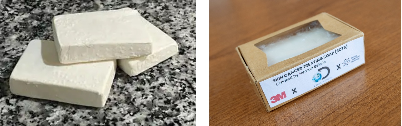
This scientist developed a soap that could help fight skin cancer. He's 14.

Link to NPR interview

A teenage boy from Fairfax, Virginia, USA, has a goal to make a soap product to fight skin cancer and be affordable. Hemen Bekele won the 2024 3M Young Scientist's Challenge in succeeding to do just that. The monetary prize of $25,000 is only the beginning.

Heman Bekele (Image from TIME magazine)

From the age of 7, Heman played around at home mixing chemicals at random, and then jump-started his learning with a chemistry set. After emigrating to the US at age 4, he recalled seeing laborers working in the sun in his home of Addis Ababa, Ethiopia, usually with no protection for their skin. His move to the U.S. sparked his thoughts about putting chemistry to use:

“When I was younger, I didn’t think much of it, but when I came to America, I realized what a big problem the sun and ultraviolet radiation is when you’re exposed to it for a long time,” Heman says. (TIME).

He read about the drug imiquimod, which is used in cream form to fight skin cancer, but he was concerned about its high cost: $40,000-$56,000 (not taking into account insurance). Because places like Ethiopia depend on agriculture for their survival, Heman thought he could help by developing something cheaper and more easily available. His idea was to create a bar of soap to deliver the drug.

Heman working at home to make the bar soap.  (tpgonlinedaily.com and Heman's website)

As long ago as 2800 BCE, Babylonians knew how to make soap, by boiling goat fat with ashes. But it was used mostly for cleaning textiles. Egyptians around 1550 BCE used their variation with animal and vegetable oils used it for personal hygiene and medicinal purposes (such as treating dermatitis, infected wounds, scalp conditions and lice. From the first century, Romans spread knowledge of soap-making across Europe, North Africa, and parts of the Middle East, but mostly used oils and scrapers. After the Roman Empire fell (476 CE), Islamic cultures--centers for science and medicine--emphasized personal hygiene due to religious practices (e.g., ritual washing). They turned soap into a daily-use item, especially for personal hygiene. From the 12th to 15th century, southern Europe created trade unions to manufacture soap, but it took until the 16th century to make it cheaply enough for common use homes.

Regular bathing—especially for personal cleanlinesswas uncommon in both Europe and America in the mid- to late-1800s. People just changed clothes or wiped their bodies. But links to disease promoted the use of soaps, as as moral, healthy, and civilized efforts.

Soap works as follows: soap molecules have a water-attracting (hydrophilic) and and a water-repelling (hydrophobic) end, and together, they bind dirt in chemical clumps and dissolve in water to wash away most things. 


Strictly speaking, the high pH of traditional soap (9-10) is not good for skin, which has a pH around 4, and it tends to dry out skin by removing oils that otherwise keep moisture in. 

The chemistry of soap wasn't known well until 1823, when Michel Eugène Chevreul showed how the Sumerian concept of soap making worked. The fat reacts with an alkali (like lye), breaking it into soap (salts of fatty acids like stearic, oleic, and palmitic acids) and glycerin. Mass production of soaps was now possible through the late 1800s, but as mentioned above, personal use was still rare. Products like Sunlight Soap (UK, 1885) and Ivory (US, 1879) then became household staples. From the 1880s on, soap was now available to the middle class.

So, a commonly used item like bar soap seems like a useful method to apply drugs to the skin, where melanomas (skin cancer) develop. Heman worked with Deborah Isabelle, a product engineering specialist at 3M, to do some computer modeling and experiments to see how chemicals added to his soap would go through the skin instead of being washed off.

Isabelle and Heman at the 3M award ceremony (NPR)

Heman's award-winning soap included organic shea butter, coconut oil and moisturizing cream 3M Cavilon. He thinks it works by reactivating a type of immune system cell called a dendritic cell, part of skin tissue, which then sends a message to the whole immune system to fight the invading cancer cells. During cancer, the dendritic cell is usually inactive, which allows the cancer to take over. If Heman's soap makes them active, the body stands a better chance to fight it. 

A mature dendritic cell with its branches (dendrites) that look like nerve cells (Wikipedia).

To go beyond a bar of soap and showing a computer model of getting drugs into the body, he needed a way to test actually doing it. At a networking event hosted by the Melanoma Research Alliance, he met Professor Vito Rebecca of Johns Hopkins University and was invited to work in his lab. 

Heman and Dr. Rebecca (Instagram)


They refined the approach from the skin absorbing a drug dissolved in it, to a specific drug carrier called a nanoparticle. If you just apply the drug in a cream or soap, it has to go through the skin randomly and wind up everywhere. But nanoparticles can be tagged to enter the skin and seek out only specific cells like the dentritic cells, where they get exposed to the drug in higher concentrations.

Left, passive random drug delivery.
Right, active focused drug delivery to a desired cell. (Pudlarz & Szemraj, 2018)


In Vito Rebecca's lab, they have tested this formulation with a form of drug called imidazoquinoline. The drug itself is not new in the war on cancer; the delivery system is. They are currently testing it on mice and, if successful, will see FDA approval in about 10 years.

Heman has described his soap as follows:

"The color is a bit of a dark type of white and it has a little bit of a bumpy texture to it, which could be a good exfoliant. It does have a strong potent medicine smell to it. But of course it isn't the worst smelling thing." (NPR)

Heman's SCTS (skin care treatment soap) in its biodegradable box (MPR News and virginiamercury.com)


Ultimately, Heman hopes to mass produce his soap and create a nonprofit organization by 2028 to distribute it worldwide. At the moment, the estimated cost for one bar of SCTS is $0.50. 

Here is a short video interview from his local TV station

Tuesday, May 6, 2025

Bioengineers made mutant, solar-powered hamster cells

Link to article

Plant cells have a structure called a chloroplast. Animal cells don't. Animal cells consume food that is provided to them after the animal eats and breaks it down into simple components. On the other hand, using chloroplasts, plants make their own food from air, water, and sunlight. But what if animal cells could be equipped with chloroplasts? And why would anyone want to do that?

You may remember from biology classes that plants use the energy from sunlight to split water molecules, which then combine with carbon dioxide to form sugars that feed the plant cells. This is called photosynthesis (photo-: light, -synthesis: building). This all takes place in the chloroplast of plant cells. A by-product of this process is oxygen, which the plant releases.

Diagram of photosynthesis (modified from Alamy)

Scientists are considering how the production of oxygen inside the body could be helpful. Here are some medical examples of when it is needed more than for just the usual supply to tissues.
  • In coronary artery disease, clogging of arteries that surround and feed the heart results in poorer blood supply and the oxygen it carries. Poor circulation often also occurs in patients with diabetes.
  • In wounds, the blood vessels are cut or crushed, so less blood with oxygen can be delivered. Body cells sent to the injury also need oxygen, but they may not get it.
  • Also, when organs or tissues suffer from trauma or disease, they need to be repaired or replaced. A special kind of science called tissue engineering can make that possible with donor cells from the body that are placed on scaffolds to grow; the structure is made inside or outside the body. Examples are small arteries, an artificial bladder, skin grafts, cartilage, and a trachea. But growing the replacement cells often suffers from insufficient oxygen.

Many methods have been used and proposed to add oxygen to areas inside the body: flooding the injury with high concentrations of oxygen, adding artificial blood substitutes that carry more oxygen, using gene therapy, or applying materials to dressings (hydrogen peroxide, nanoparticles with oxygen bubbles, nanofibers with oxygen-releasing chemicals like CaO₂, etc.). 

Some researchers have also inserted plant cells (algae) themselves!

Cyanobacteria (blue-green algae) like Synechococcus elongatus was added to rat hearts and survived 1-2 hours. The alga Chlamydomonas reinhardtii was tried on frogs and zebrafish, and the cells survived a few days. Another alga Chlorella was tested in gels or lab-grown tissues and lasted a few days to a week. 

A spongy scaffolding matrix seeded with C. reinhardtii, showing algae cells growth in the folds before implanting

Zebrafish embryo with reddish S. elongatus alga implanted in its brain

Rat heart muscle cell with S. elongatus on its surface (image from Wang et al., 2019)

One problem other than mere plant cell survival is the risk of the body's immune response attacking these cells. So, other researchers have considered inserting chloroplasts into animals cells which can be engineered to resemble the body and fool the immune response.

In 2024, researchers at Tokyo University, Waseda University, and the RIKEN Center for Sustainable Resource Science successfully implanted chloroplasts from the alga Cyanidioschyzon merolae into hamster cells. Although chloroplast implantation had been done by others earlier in different species, this was the first time photosynthetic activity was maintained in these "planimal" cells. It wasn't long, just two days, but it was still a first.

From the researchers. Blue=nuclei, magenta=chloroplasts

You might wonder why algae and their chloroplasts have been mentioned, and not higher forms of plants like leaves from flowers or trees. We think of algae as large mats of cells covering ponds and lakes, but they are made up of single-celled organisms. Each cell can exist independently. They are among the oldest of plant life on Earth, too.

Algae bloom, Lake Erie (USA), 2009 (Wikipedia)

As such, the chloroplasts in algae cells are different from those in flowering plants. Floral plant chloroplasts begin as a structure called a proplastid and then change as the plant ages from a seed to seedling to full-fledged plant. In fact, other types of plastids can come from the protoplastid and reside in different parts of the plant to produce flower colors (chromoplast), storage compartment in roots, seeds, tubers  (leucoplast), or developing leaves (etioplast, which stores material later changing to chlorophyll in chloroplasts). Gerontoplasts are old-age chloroplasts formed when plants reach the end of their life.

Image from Study.com

But chloroplasts from algae rarely change into other plastids. It might be this stability that allowed the Japanese team to succeed.

Sachihiro Matsunaga's research team first grew Cyanidioschyzon merolae in the lab, then broke open the cells chemically and physically (grinding) to release the chloroplasts. They were separated by centrifuging (spinning very fast). A heavy concentration of chloroplasts were placed in culture with hamster cells and gently shaken. These conditions probably causes a passive absorption of the chloroplasts into the cells. 

General isolation technique for chloroplasts and whole C. merolae cells (Aoki et al., 2024)

They confirmed that cells had taken up chloroplasts with fluorescent dyes and viewing under a microscope. See below from Matsunaga's study.


Next, they exposed the hamster "planimal" cells to light and measured the photosynthetic function. When they compared it to isolated chloroplasts (outside of the algae cells), they found that chloroplasts cultured with hamster cells for a day had more than half of the photosynthetic power and virtually the same after 2 days in culture. After four days, they lost the ability. The scale they used might seem small for efficiency, but regular chloroplasts in plant cells only get up to 0.6, simply because nature is not very efficient. But these results showed no loss of function after two days inside hamster cells.
 
Data modified from Aoki et al., 2024

In addition to providing medical science with cells that can produce oxygen which benefits others in the immediate vicinity, inserting chloroplasts into animal cells might have another benefit. The researchers reminded us that tissues grown in the lab could suffer from lack of oxygen because of the many layers of cells packed together. For example, it is possible to grow artificial organs, skin transplant sheets, and artificial meat in the lab.

Photo from Labiotech