How soaking in saunas could save our frogs
Link to article.
Click on images to enlarge.
There are over 7,700 species of frogs and toads worldwide. While new species are being discovered every year, many amphibians are in danger of extinction. Since the 1980s, a devastating disease has been found that is wiping them out at an alarming rate. What is this all about, and how can it be stopped?
In the 1970s and 1980s, naturalists noticed a decline in the population of many amphibian species. For example, in the U.S.:
- David Bradford (University of California) saw that only 2% of the lakes in the Siera Nevada Mountains had yellow-legged frogs in 1989 compared to surveys in 1970.
- In Oregon, 80% of the Cascade Mountains frog populations that have been monitored since the mid-1970s are gone.
- The western spotted frog in Oregon was abundant until the mid 1970s, but it is now extinct west of the Cascade Mountains from at least one third of its Oregon range of habitat. Similar stories are reported from Colorado, Arizona and New Mexico.
Dead yellow-legged frogs (Rana muscosa), Sierra Nevada Mountains, CA, 2008 (AmphibiaWeb) In other countries:
- The gastric brooding frog was discovered in 1973 north of Brisbane, Australia, and by 1981 it was thought to be extinct.
- In Costa Rica, where hundreds of golden toads used to be seen in one location in the early 1980s, fewer than a dozen were found in breeding sites less than a decade later.
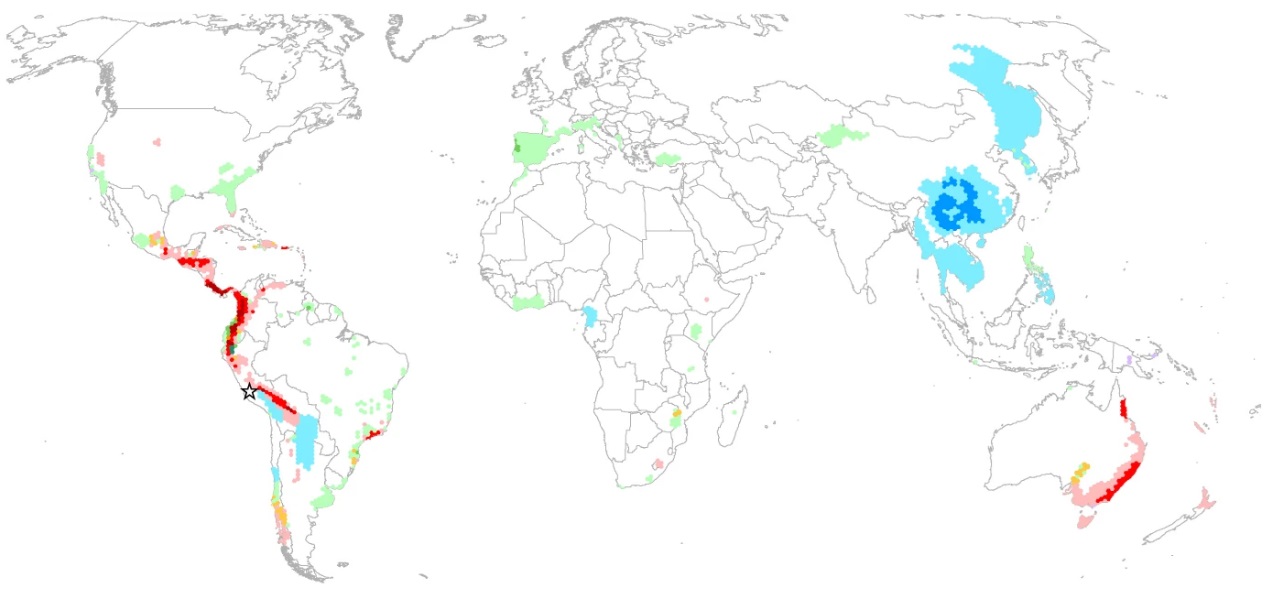
Map of amphibian declines, 1980-2004 (red=caused by diseases, primarily chytrid fungus), Nature 2023 Sometimes this happens after habitats are destroyed, but often it occurs in undisturbed areas as well. Clearly, this is a worldwide phenomenon.
This decline is important for two reasons:
- Amphibians are major consumers of invertebrates, especially insects, and of other vertebrates.
- They also serve as a food source themselves for fish, birds, mammals and aquatic insects.
In addition, amphibians are good indicators of environmental stress on land and in water, because their skin can rapidly absorb toxic chemicals. Pollutants can also affect their eggs and growth rates. Natterjack toads in Britain and tiger salamanders in the Rocky Mountains, for example, have been shown to be negatively affected by acidic conditions, not just chemicals. But by 1990, the decline couldn't always be attributed to environmental factors or human intervention. And, the more serious threats to extinction seem to be caused by diseases.
Left: types of threats to amphibians, and how many species are affected
Right: extinction level (class) bases on the type of threat (Nature, 2023) There is a fungal disease of amphibians called
chytridiomycosis,
chytrid for short (pronounced kit-rid). It is caused by two species of the fungus
Batrachochytrium (circled in red in the left graph above). The earliest occurrences were as follows:
- Titicaca water frog, Africa, 1863
- Japanese giant salamander, 1902
- African clawed frog, 1938
But those are just instances when samples from an amphibian first showed the fungus was on it, not that the fungus killed it. In the late 1990s, chytrid was determined to be the source of Australian infections. By 2004, researchers found that chytrid fungus had been found in every continent that has amphibians, except Asia. The prevalence in Africa for so many years made them feel that Africa (especially Ghana, Kenya, South Africa, and Western Africa) was the source of the infection.
One particular African frog was important for a special reason. The African clawed frog (
Xenopus laevis) became a common lab animal for
pregnancy testing in the 1930s-1940s. British zoologist
Lancelot Hogben discovered in 1930 that if frogs were injected with a pituitary gland extract from an ox, frogs would ovulate in just a few hours. The extract resembles human chorionic gonadotropin (HCG), a
hormone released by pregnant women, and he then noted that frogs ovulated when injected with their urine. This rapidly became a popular pregnancy test for several reasons:
- Results came after a few hours, not days with rabbit or mice testing with women's urine.
- Frogs could be used over and over, but mice and rabbits had to be sacrificed in order to examine their ovaries for signs of change (hence the expression "the rabbit died" to indicate a positive test).
- Maintaining frogs was easier than mice or rabbits, and the supply seemed unlimited from the African Rift Valley, as well as South Africa and Namibia.
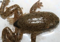
Xenopus laevis undergoing ovulation in a pregnancy test (Wikipedia) Although the Hogben frog pregnancy test has been replaced with testing that no longer involves frogs or any other animal, the
Xenopus species have since become an invasive species worldwide, presumably due to their escape in distribution or
release from labs into the environment. In fact, researchers now say that these problems in the world trade of that one frog species may have been the major reason why it spread around the world and began to infect other amphibians.
But let's get back to chytrid fungus and the way it affects amphibians.
The fungus produces spores with whiplike flagella on them to propel them through water. It then grows branches to allow it to attach to the skin, then form a sac where more spores can grow. The top contains a hole called a discharge pore where they are released.
Cytrid life cycle with actual photos of the fungus (
Yale E360)
Here is a photograph of frog skin showing actual discharge pores from chytrid spore sacs.
The first clinical signs of chytrid infection in juvenile and adult frogs may include the following symptoms:
- reddening of the underside
- abnormal spreading out of the legs
- sluggishness
- slow reflex to turn itself over when on its back
- abnormal skin shedding as white or gray material
- whitish ulcers
- spasms when handled
Gray shedding, plus splayed leg posture (Veterinary Research, 2015)
The problem is that these are symptoms usually in the last stages of chytrid growth. Early symptoms are harder to identify: anorexia and lethargy.
Fungus seems to be found on the skin in areas where keratin is present, mostly on the back, feet, and toes. Amphibian skin is vital to its existence, since they breathe through tiny lungs (except some salamanders), a lining inside their mouths, and their skin. The skin is a very thin layer of tissue that contains many blood vessels. While underwater, amphibians exchange 90-100% of their oxygen and carbon dioxide directly through their skin. This is useful short-term in warm months, but when they hibernate in winter, it is necessary to survive underwater. The skin also allows water to pass through.
As you can see from the diagram below, amphibian skin has an outer layer of epidermis (E), and directly underneath is a thick layer of capillaries (RC) to perform air and water exchange. Not shown are the vertical blood vessel branches connecting the capillary network to the arteries and veins at the bottom of the skin (LE).
RC=respiratory capillary network; E=epidermis
M=mucous gland; S and C are support tissue
SA & SV are cross-sections of arteries and veins connected vertically to the capillaries
Chytrid fungus kills amphibians by attacking the skin and producing a thickening 2-5 times thicker to 30 times thicker than normal. The normal working of the skin is reduced, so that blood electrolytes (sodium,potassium, magnesium, and chloride) decrease, and eventually, the amphibian dies of cardiac arrest, not being smothered.
Andean frog heavily infected with chytridiomycosis (New Scientist, 2019) What can be done to stop or prevent chytrid from affecting so many amphibian species? First, you have to find it, and that's not always easy.
Chytrid may be present at only 5-7 spores per liter, so detecting it in water samples requires filtering large amounts and then growing the concentrated sample on agar plates. Attempts to measure its presence on fish fins or feathers (both good sources of keratin) used as bait have proven ineffective. Free-swimming spores live for only 24 hours, and then go dormant, too, so that presents a problem in detecting them. Rock scrapings as potential attachment sites are difficult to filter. Insect bodies are not made of keratin, but an investigation into whether several types of insects might be hidden sources for chytrid have failed to find any.
Removing healthy specimens before an area is affected is time-consuming and laborious, although in some cases it has been done on a small scale. Changing the environment's chemistry or temperature is also problematic, as is adding various antifungal chemicals. Some researchers have found
a bacteria in frog skin that inhibits chytrid growth, as a sort of natural immune system. They proposed "bioaugmenting" amphibians that are in danger with this bacteria to strengthen their resistance to chytrid, and then releasing them into the wild. But this raises a lot of questions about whether this activity it may cause other problems to the ecosystem.
KM Taylor (University of California) has taken another approach in 2022. She grew the chytrid fungus on agar plates as usual, then washed the plates and filtered out the fungus and spores. What was left was any chemical the fungus had secreted. She then poured this into ponds of the Blue Oak Ranch Reserve and monitored the frogs there. Adding the chytrid metabolite increased the population of forg skin bacteria that would normally fight off chytrid. This is a promising sort of field "vaccination" still in testing.
Agar growth of chytrid; Barnett mixing up the "vaccine";
Australian researchers noted that the death rate of captive or wild frogs cut to chytrid was higher at colder temperatures. All frogs exposed to chytrid fungus in the lab at 17°C (63°F)and 23°C (73°F) died, whereas half of them at 27°C (81°F) survived. Anthony Waddle of Marquarie University (Sydney, Australia) wondered if by providing frogs with a warm place to spend time, the effect of chytrid might be prevented. So, he gave them mini-saunas.
He infected frogs with chytrid and provided them with enclosed shelters that had bricks with holes to serve as miniaure saunas. Frogs could choose between shaded and unshaded areas, with or without saunas. Waddle said, "
We found frogs flocked to the sunny saunas, heated up their little bodies, and quickly fought off infection." The saunas were built with easily accessible materials: plastic greenhouse shelter, masonry bricks, plastic cable ties, and black paint. Details can be found
online here.
Distribution of frog saunas in moist ground near a water source.
It is not clear yet what exactly kills the chytrid fungus. The frogs that Waddle tested were shown to rise in body temperature to 30°C (86°F), which he said
"is sufficient to kill chytrid fungi". But is it the temperature itself, or does the warming somehow enhance the bacteria that produce chemicals against it? More research is needed, but for now, at least these inexpensive frog saunas can be used in areas like parks or other natural habitats that are not hard to reach.


No comments:
Post a Comment